Only about one in 5,000 drugs developed to treat cancer proves viable for clinical use, according to WSU Elson S. Floyd College of Medicine Associate Professor Weimin Li, MD, PhD. And behind that one in 5,000 is decades of work from scientists in fields ranging from the foundational to the applied.
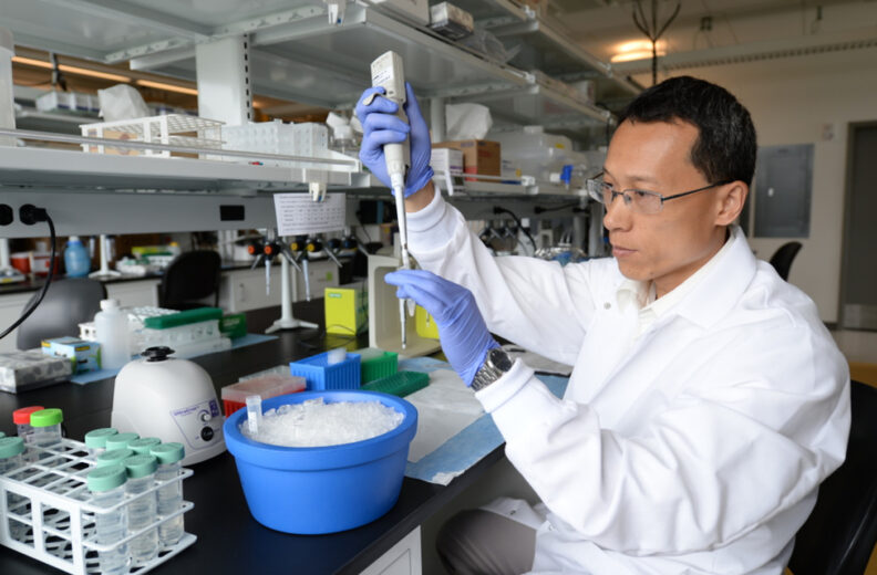
Li is one of those scientists working to improve clinical cancer detection and treatment by uncovering how cancers function at a molecular level. This World Cancer Day, he’s pulling back the curtain on his work in the college’s Department of Translational Medicine and Physiology and findings he and his team hope to translate from the lab into practice to help patients and health care providers in Washington and beyond.
Most cancer researchers specialize in studying one small piece of the complex puzzle of cancer. For Li, spurred to enter research after a career as a practicing oncologist, that piece of the puzzle is creating more effective laboratory models to better understand breast cancer progression and control.
“We still cannot conquer this disease with current therapeutic tools,” Li said. “In-depth study at the molecular, biochemical level could solve some fundamental questions.”
In order to identify better biomarkers for cancer detection and treatment, Li and his team created a unique 3D tissue culture model for breast cancer that realistically recreates the conditions in which tumors grow in the human body. The work is supported by funding from WSU’s Office of Commercialization and several external grants.
Using an accurate model in lab testing is essential, Li said. His team has published several studies showing differences in how realistically existing models perform, and their model has been recognized and adopted by other research labs around the country.
Li’s model is unique because it was created with a native tissue-derived extracellular matrix, with human breast cancer cells grown in an environment that closely resemble conditions in the human body. All cells in the body are surrounded by material referred to as the extracellular matrix (ECM). Cancer cells modify the ECM and other nearby material, called the tumor microenvironment, to promote their growth and spread to other tissues. Understanding that ECM remodeling process is key to understanding and preventing cancer progression.
Li and his team use their 3D culture model to identify and validate biomarkers, or specific molecules, to target in better diagnostic tests and treatments for breast cancer. Their technology enables better detection, more accurate prognoses, and more effective treatments, achieving better outcomes for patients.
“We are excited every day, quite frankly, by our findings,” Li said.
The team is working on identifying biomarkers that predict which breast cancer patients are at risk of their cancer spreading, in particular patients with ductal carcinoma in situ (DCIS).
DCIS is sometimes called “stage 0” breast cancer because the cancer cells are slow-growing and in most cases will never spread outside of one milk duct. Given the low probability of metastasis, some oncologists feel that invasive treatments like radiation therapy or a mastectomy do more harm than good in DCIS. But how can doctors know whether a particular patient’s cancer will spread or not?
Li and his team are collaborating with Washington clinicians to answer that question. Using their model, the team’s goal is to identify biomarkers that predict the risk of a DCIS patient’s cancer spreading.
“If our predictions prove to be valid,” Li said, “this can be translated directly into clinical practice.”